🫀
Renal · Anatomy
Urinary System
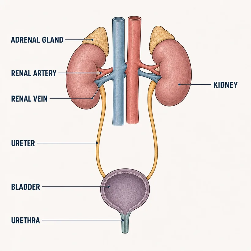
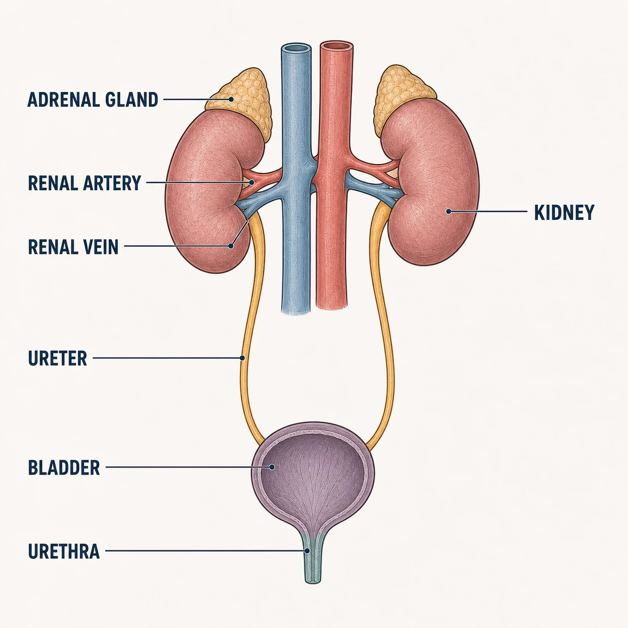
Kidneys make urine; ureters carry it; the bladder stores it; the urethra drains it.
The structures
- Kidneys — Retroperitoneal, T12–L3; left sits slightly higher
- Renal artery & vein — A fifth of cardiac output flows through every minute
- Ureters — Peristaltic tubes — stones lodge at their three narrow points
- Bladder — Detrusor muscle; ~400–600 mL comfortable capacity
- Urethra — Short in women (UTI risk), long in men (retention risk)
- Adrenal glands — Sit on top of each kidney — different organ, shared real estate
Why it matters on shift
- Flank pain radiating to the groin = classic ureteral stone — strain the urine
- Sudden agitation in an elderly patient? Palpate for a distended bladder before reaching for sedation
- Costovertebral angle tenderness points to kidney (pyelonephritis), not bladder
Source: Anatomical structures per OpenStax Anatomy & Physiology, 2nd ed. (openstax.org, CC BY 4.0). Educational reference — verify clinical application against your facility's policy.Reference only — verify against your facility's policy.