🫀
Respiratory · Anatomy
Lungs & Airway
Air travels trachea → bronchi → bronchioles → alveoli, where gas exchange happens.
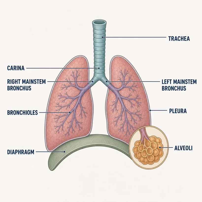
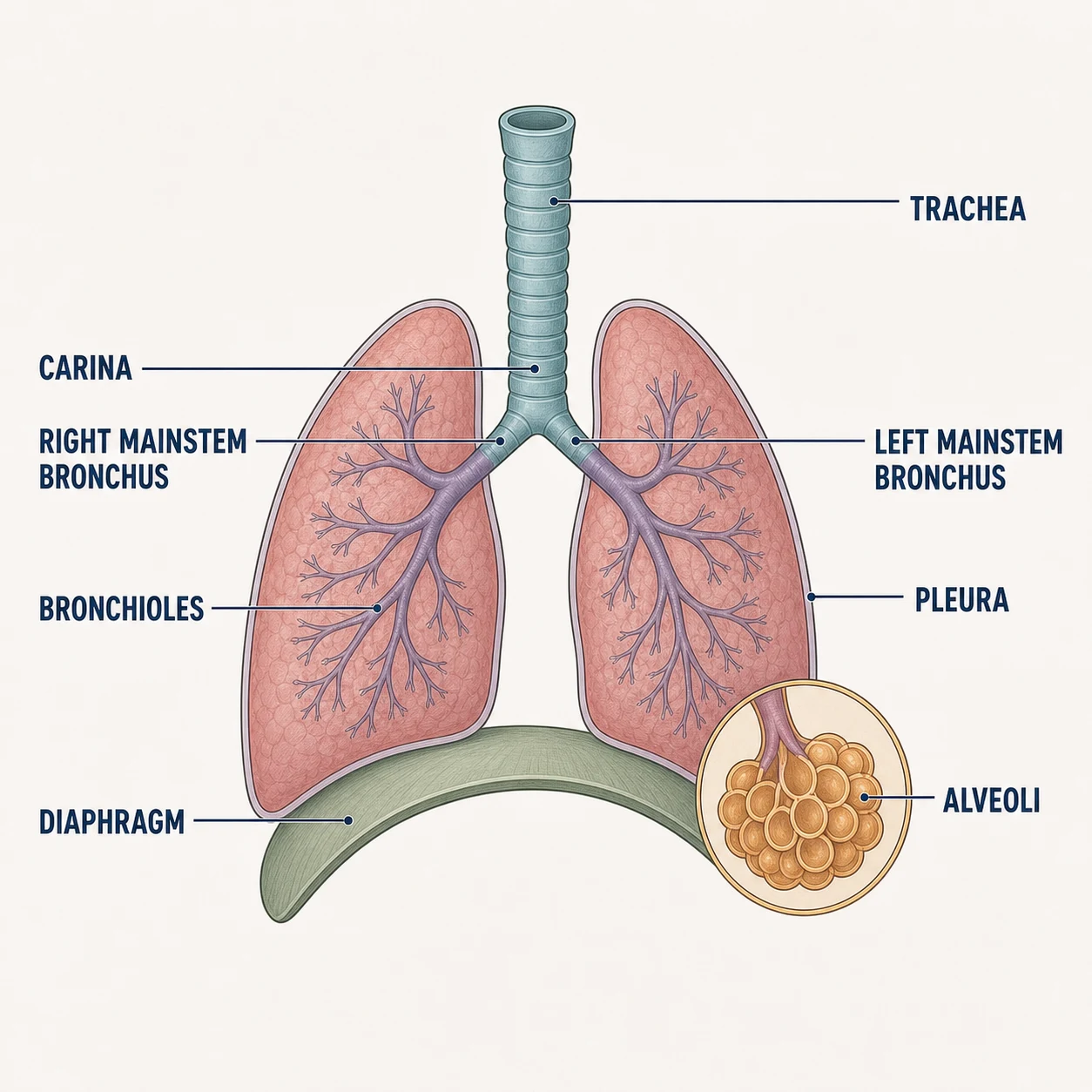
The structures
- Trachea — Rigid airway held open by C-shaped cartilage rings
- Carina — Split into right and left mainstem bronchi — suction trigger point
- Right mainstem bronchus — Wider and more vertical — where aspirated objects and deep ET tubes go
- Bronchioles — Smooth-muscle airways — the site of bronchospasm
- Alveoli — Gas-exchange sacs wrapped in capillaries
- Pleura — Two membranes with a potential space — where effusions and pneumothorax live
- Diaphragm — Primary muscle of breathing (C3–C5, phrenic nerve)
Why it matters on shift
- ET tube advanced too far almost always ends up in the RIGHT mainstem — breath sounds vanish on the left
- Crackles = fluid in alveoli; wheeze = tight bronchioles; stridor = upper airway — three different emergencies
- C3–C5 keeps the diaphragm alive: high cervical injuries threaten breathing
Source: Anatomical structures per OpenStax Anatomy & Physiology, 2nd ed. (openstax.org, CC BY 4.0). Educational reference — verify clinical application against your facility's policy.Reference only — verify against your facility's policy.