🫀
Cardiovascular · Anatomy
Heart
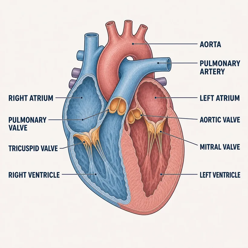
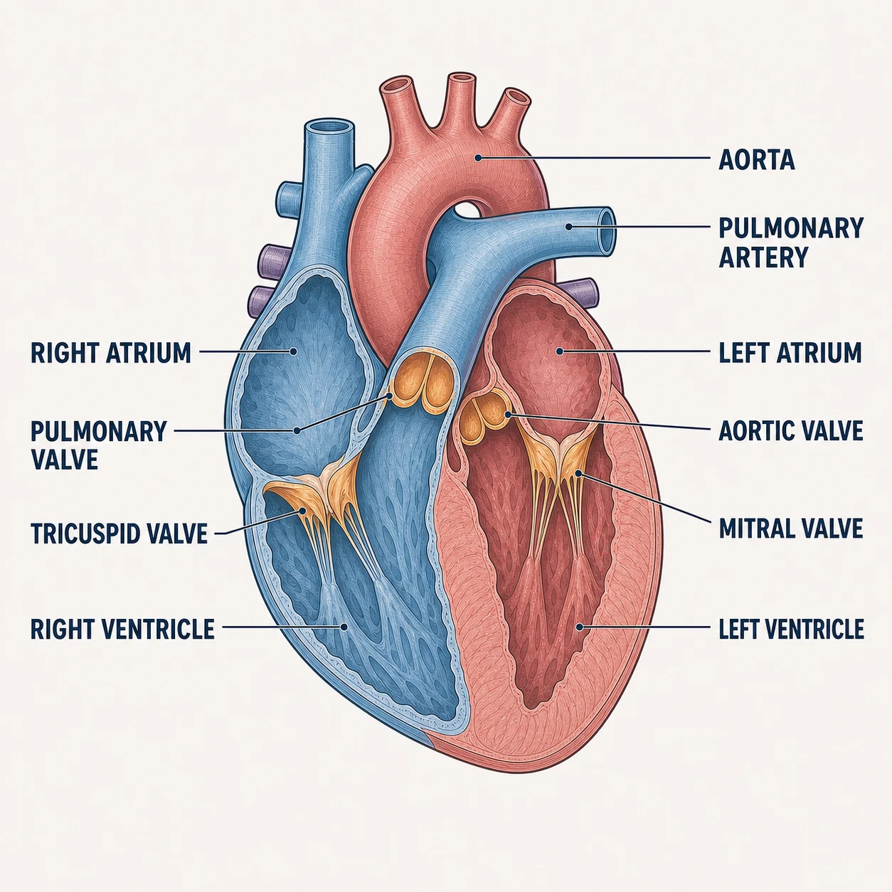
Four chambers, four valves, one loop: body → right heart → lungs → left heart → body.
The structures
- Right atrium — Receives deoxygenated blood from the vena cavae
- Tricuspid valve — Right atrium → right ventricle
- Right ventricle — Pumps to the lungs via the pulmonary artery
- Pulmonary valve — Right ventricle → pulmonary artery
- Left atrium — Receives oxygenated blood from the pulmonary veins
- Mitral valve — Left atrium → left ventricle
- Left ventricle — Thickest wall — pumps to the whole body via the aorta
- Aortic valve — Left ventricle → aorta
Why it matters on shift
- Murmur = a valve problem until proven otherwise; mitral and aortic are the ones you'll hear about most
- Left-sided failure backs up into the LUNGS (crackles, dyspnea); right-sided backs up into the BODY (JVD, edema)
- MI territory matters: an inferior MI (RCA) can drop preload — think twice before nitro
Source: Anatomical structures per OpenStax Anatomy & Physiology, 2nd ed. (openstax.org, CC BY 4.0). Educational reference — verify clinical application against your facility's policy.Reference only — verify against your facility's policy.