🫀
Gastrointestinal · Anatomy
GI Tract
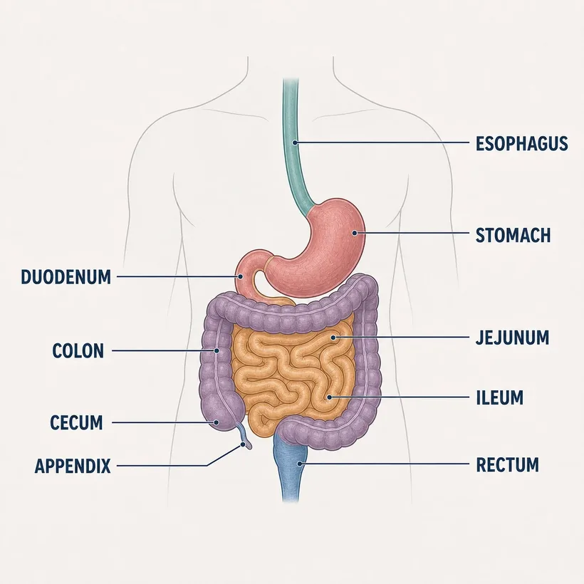
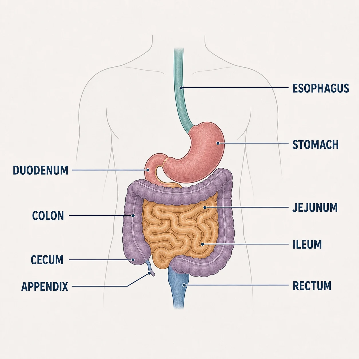
One continuous tube: mouth → esophagus → stomach → small intestine → colon → rectum.
The structures
- Esophagus — Peristalsis carries food past the diaphragm's hiatus
- Stomach — Acid + churning; empties into the duodenum
- Duodenum — First stop for bile and pancreatic enzymes — most ulcers here
- Jejunum & ileum — Where nearly all absorption happens
- Cecum & appendix — RLQ landmark — McBurney's point
- Colon — Water reabsorption; ascending → transverse → descending → sigmoid
- Rectum — Storage and the defecation reflex
Why it matters on shift
- Bowel sounds absent + distension + pain = obstruction workup, not 'wait and see'
- Upper GI bleed = coffee-ground emesis or melena; lower = bright red — different sources, different urgency
- NG tube placement is verified by X-ray, not by auscultating air — that habit has killed patients
Source: Anatomical structures per OpenStax Anatomy & Physiology, 2nd ed. (openstax.org, CC BY 4.0). Educational reference — verify clinical application against your facility's policy.Reference only — verify against your facility's policy.